Whole-Brain Calcium Imaging during Physiological Vestibular Stimulation in Larval Zebrafish
G. Migault
,
T.L. van der Plas
,
H. Trentesaux
,
T. Panier
,
R. Candelier
,
R. Proville
,
B. Englitz
,
G. Debrégeas
,
V. Bormuth
Current Biology, 28, 1-13
Published 15 Nov. 2018
DOI: https://doi.org/10.1016/j.cub.2018.10.017
ISSN: 0960-9822
Abstract
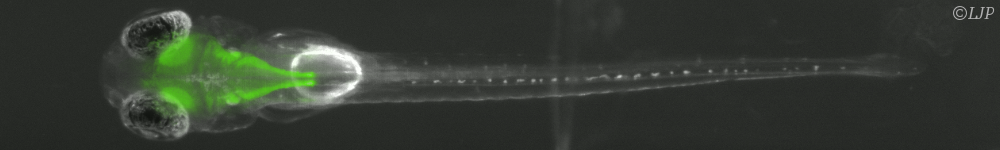
The vestibular apparatus provides animals with postural and movement-related information that is essential to adequately execute numerous sensorimotor tasks. In order to activate this sensory system in a physiological manner, one needs to macroscopically rotate or translate the animal?s head, which in turn renders simultaneous neural recordings highly challenging. Here we report on a novel miniaturized, light-sheet microscope that can be dynamically co-rotated with a head-restrained zebrafish larva, enabling controlled vestibular stimulation. The mechanical rigidity of the microscope allows one to perform whole-brain functional imaging with state-of-the-art resolution and signal-to-noise ratio while imposing up to 25° in angular position and 6,000°/s^2 in rotational acceleration. We illustrate the potential of this novel setup by producing the first whole-brain response maps to sinusoidal and stepwise vestibular stimulation. The responsive population spans multiple brain areas and displays bilateral symmetry, and its organization is highly stereotypic across individuals. Using Fourier and regression analysis, we identified three major functional clusters that exhibit well-defined phasic and tonic response patterns to vestibular stimulation. Our rotatable light-sheet microscope provides a unique tool for systematically studying vestibular processing in the vertebrate brain and extends the potential of virtual-reality systems to explore complex multisensory and motor integration during simulated 3D navigation.
Cette publication est associée à :
Imagerie calcique et comportement du poisson zèbre et Danionella Cerebrum