
|
Multiphoton imaging of developing tissues: multimodal, multicolor and light-sheet approaches
Par Emmanuel Beaurepaire & Willy Supatto
Le 28 Juin 2016 à 11h00 - Seminar room LJP (tower 32-33, 5th floor)
|
Résumé
Modern issues in systems biology require tissue-scale measurements of multiple cell parameters. Multiphoton fluorescence microscopy has proven invaluable for tissue studies with its ability to provide subcellular resolution in thick/live samples. However in practice, current methods are still limited in terms of speed, depth, innocuity, and ability to simultaneously probe multiple parameters.
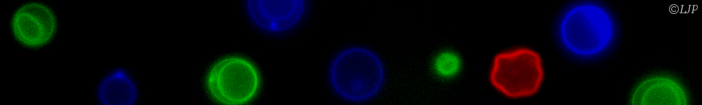
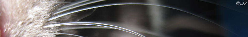
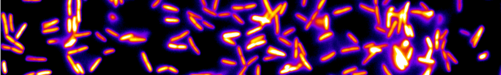
In this presentation we will first introduce the principles and stakes of this field at the interface between physics and biology. We will then discuss some ongoing developments aiming at adressing the issues mentionned above: efficient combination of fluorescence with harmonics contrasts [1,2], multicolor two-photon excitation [3,4], and parallelization through light-sheet excitation [5-7]. We will illustrate the benefit of these developments for applications in developmental biology (embryogenesis, brain development) and for biomedical applications (imaging of optically accessible tissues such as the eye and skin).
References:
[1] Olivier, Science (2010). [2] Dray, Development (2015). [3] Mahou, Nat Methods (2012). [4] Loulier, Neuron (2014). [5] Truong, Nat Methods (2011). [6] Mahou, Nat Methods (2014). [7] Wolf, Nat Methods (2015).