Label free multiphoton imaging of human pulmonary tissues through two-meter-long microstructured fiber and multicore image-guide
G. Ducourthial
,
C. Lefort
,
D.A. Peyrot
,
T. Mansuryan
,
S.G. Kruglik
,
C. Vever-Bizet
,
L. Thiberville
,
F. Lacombe
,
G. Bourg-Heckly
,
F. Louradour
ENDOSCOPIC MICROSCOPY VIII, 8575, 85750H (2013)
DOI: 10.1117/12.2003118
ISSN: 0277-786X
Abstract
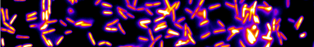
This work deals with label free multiphoton imaging of the human lung tissue extra-cellular matrix (ECM) through optical fibers. Two devices were developed, the first one using distal scanning associated to a double clad large mode area (LMA) air-silica microstructured fiber, the second one using proximal scanning of a miniature multicore image guide (30000 cores inside a 0.8 mm diameter). In both cases, the main issue has been efficient linear and nonlinear distortion pre-compensation of excitation pulses. By inserting before the delivery fiber a compact (10 cm x 10 cm footprint) grisms-based stretcher (a grating in close contact with a prism) made of readily available commercial components, we achieved as short as 35-femtosecond-duration pulses that were temporally compressed at the direct exit of a 2-meter-long fiber. Interestingly, this femtosecond pulse fiber delivery device is also wavelength tunable over more than 100 nm inside the Ti: Sapphire emission band. With the help of distal scan system, those unique features allowed us to record elastin (through two-photon fluorescence) and collagen (through second harmonic generation) fibered network images. These images were obtained ex-vivo with only 15 mW @ 80 MHz of IR radiation delivered to the alveoli or bronchus tissues. 3D imaging with 400-m-penetration depth inside the tissue was possible working with a 2-meter-long LMA fiber. With the help of proximal scanning, the miniature image guide allowed us to perform endoscopic real time microimaging of the ECM ex vivo.
Cette publication est associée à :
Cette publication est associée à :